Mouse B2m Knockout Cell Line-MC38
Cat.No. : CSC-RT2755
Host Cell: MC38 Target Gene: B2m
Size: 1x10^6 cells/vial, 1mL Validation: Sequencing
Cat.No. : CSC-RT2755
Host Cell: MC38 Target Gene: B2m
Size: 1x10^6 cells/vial, 1mL Validation: Sequencing
| Cat. No. | CSC-RT2755 |
| Cell Line Information | This cell is a stable cell line with a homozygous knockout of mouse B2m using CRISPR/Cas9. |
| Target Gene | B2m |
| Host Cell | MC38 |
| Size Form | 1 vial (>10^6 cell/vial) |
| Shipping | Dry ice package |
| Storage | Liquid Nitrogen |
| Species | Mus musculus (Mouse) |
| Revival | Rapidly thaw cells in a 37°C water bath. Transfer contents into a tube containing pre-warmed media. Centrifuge cells and seed into a 25 cm2 flask containing pre-warmed media. |
| Mycoplasma | Negative |
| Format | One frozen vial containing millions of cells |
| Storage | Liquid nitrogen |
| Safety Considerations |
The following safety precautions should be observed. 1. Use pipette aids to prevent ingestion and keep aerosols down to a minimum. 2. No eating, drinking or smoking while handling the stable line. 3. Wash hands after handling the stable line and before leaving the lab. 4. Decontaminate work surface with disinfectant or 70% ethanol before and after working with stable cells. 5. All waste should be considered hazardous. 6. Dispose of all liquid waste after each experiment and treat with bleach. |
| Ship | Dry ice |
Defective MHC class I antigen presentation is considered the most common mechanism of cancer immune escape. Despite its increasing prevalence, its mechanistic implications and potential strategies to address this challenge remain poorly understood. By studying a mouse tumor model deficient in β2-microglobulin (B2M), researchers found that MHC class I loss leads to immune desertification of the tumor microenvironment (TME) and broad therapeutic resistance to immunotherapy, chemotherapy, and radiotherapy. The study demonstrated that treatment with long-acting mRNA-encoded interleukin 2 (IL2) restored immune cell-infiltrating, IFNγ-promoting, highly pro-inflammatory TME features and, when combined with a tumor-targeting monoclonal antibody (mAb), could overcome therapeutic resistance. Surprisingly, the effectiveness of this treatment was driven by neoantigen-specific IFNγ-releasing CD8+ T cells that recognized neoantigens cross-presented by TME-resident activated macrophages that acquired enhanced antigen presentation capacity and other M1 phenotype-associated features under IL2 treatment. These findings highlight the unexpected importance of restoring neoantigen-specific immune responses in treating MHC class I-deficient cancers.
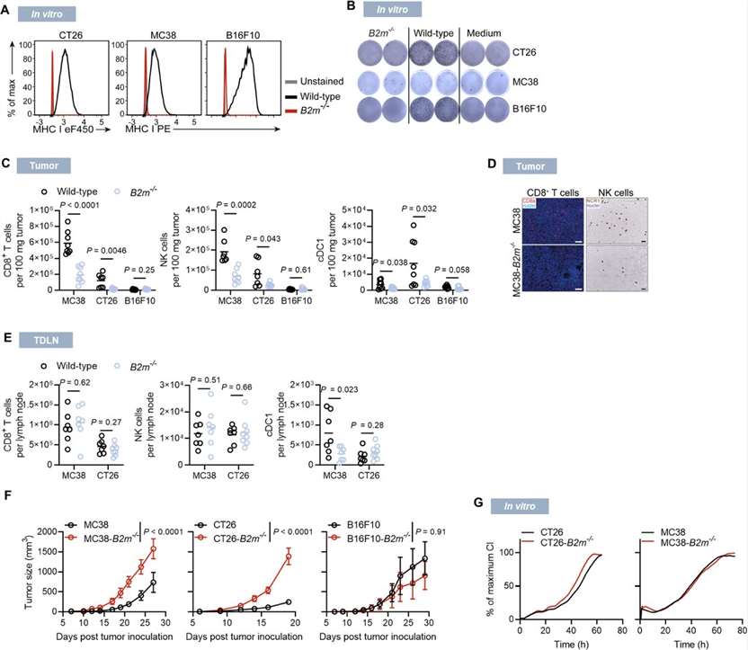
B2M is a common component of all MHC class I molecules. Its loss results in a complete loss of MHC class I surface expression, resulting in a loss of CD8+ T cell recognition. To investigate MHC class I presentation defects in different settings, the researchers used three mouse B2m knockout tumor cells: CT26, MC38, and B16F10. Among them, CT26 and MC38 form highly immunogenic tumors and trigger spontaneous CD8+ T cell responses, while B16F10 melanoma has a low prevalence of tumor-infiltrating leukocytes (TILs) and is considered a non-immunogenic tumor. B2m knockout (B2m-/-) cells lack MHC class I surface expression and therefore cannot be recognized by co-cultured antigen-specific CD8+ T cells (Figure 1A and B). In syngeneic mice, longitudinal analysis of immune cell infiltration during the growth of wild-type and B2m-/- tumors revealed a gradual reduction in immune cell infiltration in CT26-B2m-/- tumors, with immune cell desertification of the tumor microenvironment (TME) within 20 days after inoculation, and a reduction in CD8+ T cells, NK cells, and conventional type I dendritic cells (cDC1) (Figure 1C). B2M knockout had similar effects on MC38 tumors, while B16F10 tumors, regardless of their B2m genotype, showed sparse immune infiltration (Figures 1C and D). CT26-B2m knockout and MC38-B2m knockout tumors showed significantly faster progression in vivo, but not in in vitro cell culture conditions (Figures 1F and G), while B16F10 tumors showed no significant growth differences compared with wild-type tumors (Figure 1F).
 Figure 1. Characterization of MHC class I-deficient tumors. (Beck J D, et al., 2023)

Our promise to you:
Guaranteed product quality, expert customer support.
 24x7 CUSTOMER SERVICE
 CONTACT US TO ORDER